Anatomy of the foot
Structure & fonctionnement du pied
- Publié le 10 Janvier 2020
- Update on
- By Forlini Orthopedie
SUMMARY :
The human foot is a complex anatomical structure. composed of 26 bones, around 30 joints, and more than 100 muscles, tendons, and ligaments. Il constitue la base de l’appui et joue un rôle fondamental dans l’équilibre, le déplacement, et l’absorption des chocs. Comprendre l’anatomie du pied permet de mieux identifier l’origine des douleurs plantaires et de proposer des solutions adaptées pour soutenir et rééquilibrer la structure du pied.
Foot skeleton
The foot is divided into three main anatomical parts:
The hindfoot
- Talus (or Astragalus): It articulates with the tibia and fibula to form the ankle joint (talocrural joint).
- Calcaneus (or Calcaneum): From the heel, the largest bone of the foot, it supports the body's weight and receives the insertions of the Achilles tendon.
The midfoot
- Navicular (or scaphoid)
- Cuboid
- Three cuneiforms (medial, intermediate, lateral)
These bones form the arch of the foot and play a role in shock absorption and propulsion.
The forefoot
- Metatarsals (5 long axles)
- Phalanges (14 bones): Each toe has 3 (proximal, middle, distal), except for the big toe (hallux), which has 2.
Les trois types morphologiques de pieds
On distingue classiquement trois morphologies de pieds, déterminées par la longueur relative des orteils. Cette classification a des implications sur le chaussage et la prédisposition à certaines pathologies.
Le pied égyptien
Présent chez environ 50 % de la population, c’est la morphologie la plus répandue. Le gros orteil (hallux) est le plus long, les orteils suivants décroissant progressivement en longueur. Ce type de pied est plus exposé au risque d’Hallux valgus (oignon du gros orteil), car la forme en pente favorise la compression de l’avant-pied dans les chaussures étroites.
Le pied grec
Observé chez 23 % des individus, il se caractérise par un deuxième orteil plus long que le gros orteil. Cette configuration, souvent associée à la statuaire grecque antique, est la plus facile à chausser car elle épouse naturellement la forme pointue de la plupart des souliers manufacturés. Elle peut néanmoins favoriser une Metatarsalgia (douleur sous l’avant-pied) en raison d’un appui majoré sur la tête du deuxième métatarsien.
Le pied romain ou carré
Concernant 27 % de la population, le pied romain présente un alignement des trois premiers orteils, qui sont de longueur sensiblement égale. Cette morphologie offre une répartition homogène des appuis sur l’avant-pied, ce qui en fait un pied peu sujet aux déformations liées au chaussage. Il peut toutefois être associé à un flat foot en raison d’une surface d’appui antérieure plus large.


Articulations et mobilité du pied
The foot contains about thirty joints, the main ones being:
- Subtalar joint: between the talus and calcaneus, allows for inversion and eversion movements.
- Chopard's Articulation complex articulation located between the hindfoot and the midfoot. It unites two bones of the hindfoot (the talus and calcaneus) with two bones of the midfoot (the navicular and cuboid). It allows for pronation/supination movements, contributes to the mobility of the plantar arch, and acts as a dynamic shock absorber during walking and running. It can be «locked» by certain movements (supination), strengthening the rigidity of the foot during the propulsive phase.
- Tarsometatarsal Joints (Lisfranc's Joints): ensure arch flexibility.
- Metatarsophalangeal joints important for the progression of the stride and propulsion during walking. When these joints collapse or cause pain, we speak of metatarsalgias.
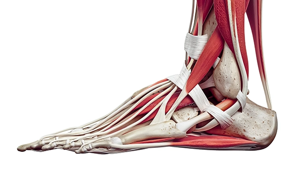
Ligaments and stability
Among the many ligaments of the foot, some ensure joint stability and maintain the arch of the foot:
- Plantar calcaneonavicular ligament (or «spring ligament») medial longitudinal arch.
- Plantar fasciitis a tense fibrous structure extending from the calcaneus to the toes, essential in plantar fasciitis. When the aponeurosis is inflamed, it is called’Plantar fasciitis.
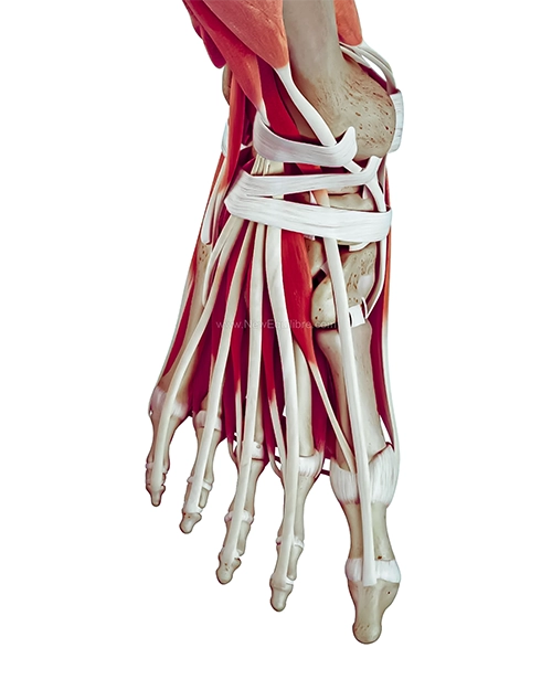
Principaux muscles et tendons du pied
The muscles of the foot are divided into intrinsic muscles (entirely within the foot) and extrinsic muscles (originating in the leg).
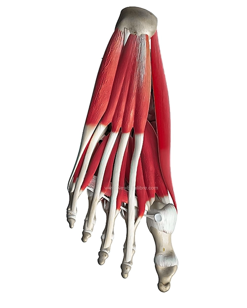
Muscles intrinsèques du pied
Les muscles intrinsèques prennent leur origine et se terminent dans le pied. Ils assurent la stabilité fine des orteils et participent au maintien des arches plantaires. On les regroupe en quatre couches anatomiques.
| Muscle | Action principale | Innervation |
|---|---|---|
| Couche superficielle (1ʳᵉ couche) | ||
| Abducteur de l’hallux | Abduction du gros orteil | Nerf plantaire médial |
| Court fléchisseur des orteils | Flexion des orteils II à V | Nerf plantaire médial |
| Abducteur du Vᵉ orteil | Abduction du 5ᵉ orteil | Nerf plantaire latéral |
| 2ᵉ couche | ||
| Carré plantaire | Assiste la flexion des orteils | Nerf plantaire latéral |
| Lombricaux (×4) | Flexion métatarsophalangienne + extension interphalangienne | Nerfs plantaires médial et latéral |
| 3ᵉ couche | ||
| Court fléchisseur de l’hallux | Flexion du gros orteil | Nerf plantaire médial |
| Adducteur de l’hallux | Adduction du gros orteil | Nerf plantaire latéral |
| Court fléchisseur du Vᵉ orteil | Flexion du 5ᵉ orteil | Nerf plantaire latéral |
| 4ᵉ couche (interosseux) | ||
| Interosseux dorsaux (×4) | Abduction des orteils II à IV | Nerf plantaire latéral |
| Interosseux plantaires (×3) | Adduction des orteils III à V | Nerf plantaire latéral |
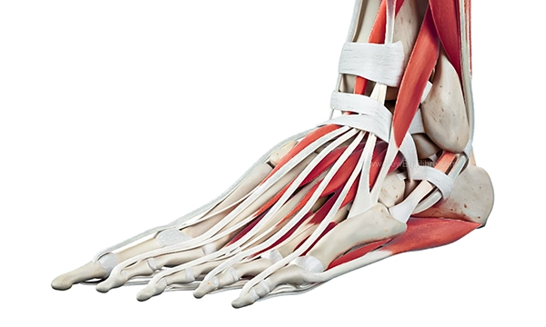
Muscles extrinsèques et tendons principaux
Les muscles extrinsèques prennent leur origine dans la jambe et leurs tendons traversent la cheville pour s’insérer dans le pied :
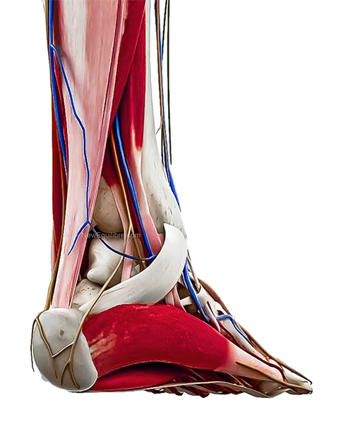
- Achilles tendon (triceps sural) : insertion sur le calcanéus, moteur de la flexion plantaire de la cheville. Indispensable à la marche, la course et le saut.
- Tendon tibial postérieur : soutien de la voûte plantaire et inversion du pied.
- Tendon tibial antérieur : flexion dorsale de la cheville (relève le pied).
- Tendons fibulaires (long et court) : éversion du pied et stabilisation latérale de la cheville.
Vascularization and innervation
Vascularisation : assurée par les artères plantaires (médiale et latérale) issues de l’artère tibiale postérieure.
Innervation : dépend des nerfs tibial, fibulaire profond et superficiel, et sural.
Innervation du pied
Le pied est innervé par plusieurs branches nerveuses issues du nerf sciatique, qui se divise au niveau du genou :
- Nerf tibial : innerve la plante du pied via ses deux branches terminales, le nerf plantaire médial et le nerf plantaire latéral. Il commande la majorité des muscles intrinsèques et assure la sensibilité de la voûte plantaire. Sa compression dans le canal tarsien (en arrière de la malléole interne) provoque le syndrome du tunnel tarsien, responsable de douleurs et de fourmillements plantaires.
- Nerf fibulaire superficiel : innerve la face dorsale du pied et les muscles fibulaires.
- Nerf fibulaire profond : innerve le muscle court extenseur des orteils et la sensibilité de la première commissure interdigitale (entre le gros orteil et le 2ᵉ orteil).
- Nerf saphène (branche du nerf fémoral) : innerve la face médiale du pied.
- Nerf sural : innerve la face latérale du pied et le bord externe.
Vascularisation
La vascularisation artérielle du pied repose sur deux axes principaux :
- L’artère dorsale du pied (pédieuse), branche de l’artère tibiale antérieure, qui parcourt la face dorsale.
- The artères plantaires médiale et latérale, branches de l’artère tibiale postérieure, qui irriguent la plante du pied et forment l’arcade plantaire profonde.
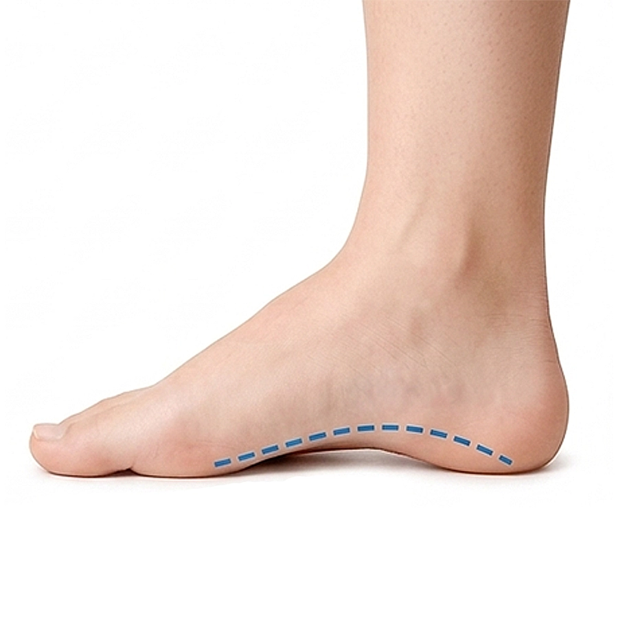
Foot arches
Le pied humain comporte trois voûtes plantaires :
- Medial arch the highest, the most biomechanically important.
- Outer arch plus plate, stabilizer.
- Anterior arch passes through the metatarsal heads.
These arches allow for shock absorption, pressure redistribution, and foot propulsion during walking or running.
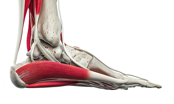
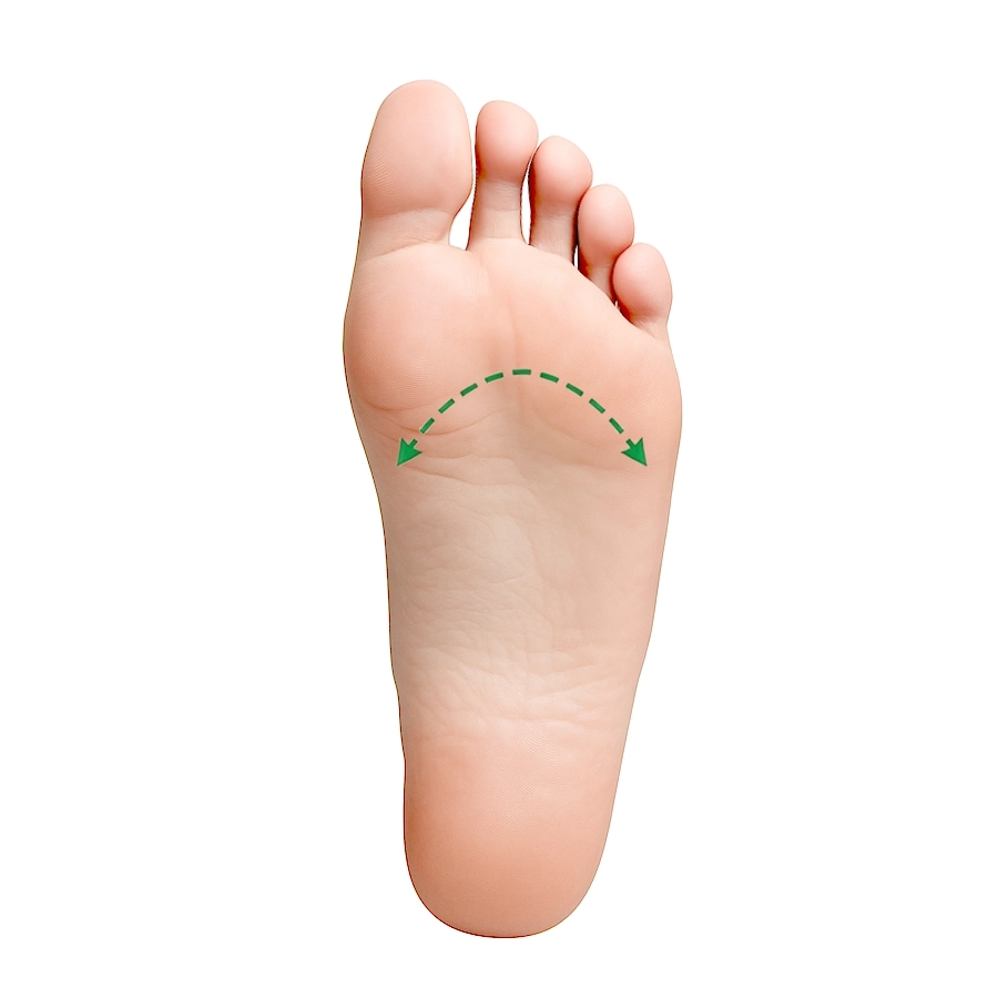
Le fascia plantaire (aponévrose plantaire)
The fascia plantaire est une bande épaisse de tissu conjonctif qui s’étend du calcanéus (os du talon) jusqu’à la base des orteils. Véritable tendeur de la voûte plantaire, il joue un rôle essentiel dans :
- The soutien de l’arche longitudinale médiale du pied
- L’absorption des chocs lors de l’impact au sol
- The transmission des forces entre l’arrière-pied et l’avant-pied pendant la marche
Lorsque le fascia plantaire est soumis à des tensions excessives — par exemple en cas de High-arched foot, de excessive pronation, ou de station debout prolongée — il peut s’enflammer à son insertion calcanéenne. On parle alors de plantar fasciitis (ou aponévrosite plantaire), l’une des causes les plus fréquentes de douleur au talon.

Foot biomechanics
The foot undergoes forces equivalent to several times the body's weight with each step. It functions according to a biomechanical triptych:
- Tardigrade phase Heel support
- Digitigrade Midfoot stabilization
- Digitigrade phase Forefoot propulsion
Un déséquilibre dans une de ces phases d'appuis du pied peut entraîner :
- Pain (plantar fasciitis, metatarsalgias, Talalgias…),
- Chronic pathologies (Hallux valgus, calcaneal spur…),
- Postural problems (knees, hips, lower back...).
Pour aller plus loin, découvrez notre guide complet sur la biomécanique du pied :
Link between foot anatomy and pathologies
Les semelles orthopédiques permettent de soutenir les appuis du pied et d’ainsi rééquilibrer l’ensemble de la posture. Nos orthopédistes du cabinet Forlini ont conçus les semelles New Equilibre, accessibles en quelques clics par notre boutique en ligne, pour offrir à tous du confort au quotidien. Leur structure thermoformée adaptative se moule à votre pied dès les premieres utilisation pour s’adapter à votre morphologie. Chaque modèle de semelles New Equilibre est conçu avec des matériaux spécifiques à chaque activité, pour le quotidien, le travail et le sport.
Rééquilibrer les appuis du pied pour soulager les douleurs
Understanding anatomical structures allows for the identification of the origin of many pathologies. Thus, we were able to design New Equilibre insoles. Made in France, our orthopedic insoles are based on our over 35 years of expertise in orthopedic practice. We offer specific models for the most common pathologies to relieve and prevent pain:
Optimisez votre confort et vos performances sportives ! À chaque sport ses semelles pour une efficacité optimale.
FAQ - Les questions fréquemment posées sur le pied:
Combien d'os y a-t-il dans le pied humain ?
Le pied humain adulte compte 26 os : 7 os du tarse (calcanéus, talus, cuboïde, naviculaire, 3 cunéiformes), 5 métatarsiens et 14 phalanges. Soit 52 os pour les deux pieds, ce qui représente un quart des 206 os du corps humain.
Quels sont les trois types de pieds ?
On distingue le pied égyptien (gros orteil le plus long, 50 % de la population), le pied grec (2ᵉ orteil plus long que le gros orteil, 23 %) et le pied romain ou carré (trois premiers orteils alignés, 27 %). Chaque type a des implications spécifiques sur le chaussage et les risques pathologiques.
C'est quoi le fascia plantaire ?
Le fascia plantaire (ou aponévrose plantaire) est une bande épaisse de tissu conjonctif qui relie le calcanéus à la base des orteils. Il soutient la voûte plantaire, absorbe les chocs et transmet les forces lors de la marche. Son inflammation est responsable de la plantar fasciitis.
Quelle est la différence entre pronation et supination ?
The pronation est l’inclinaison du pied vers l’intérieur lors de la marche ou de la course. La supination est l’inclinaison vers l’extérieur. Un excès de pronation peut entraîner une fasciite plantaire ou un syndrome de Morton ; un excès de supination favorise les entorses de cheville et les fractures de stress.
Comment s'appellent les parties du pied ?
Le pied se divise en trois régions : l’arrière-pied (calcanéus, talus), le médio-pied (naviculaire, cuboïde, cunéiformes) et l’forefoot (métatarsiens, phalanges). On distingue également la face dorsale (dessus du pied) et la face plantaire (plante du pied).
Article rédigé par Forlini Orthopedie
Spécialiste de la fabrication de semelles orthopédiques
- Plus de 35 ans d’expérience dans la prise en charge du pied (depuis 1988)
- Plus de 5000 patients accompagnés chaque année en cabinet.
- À l’origine du développement des semelles New Equilibre